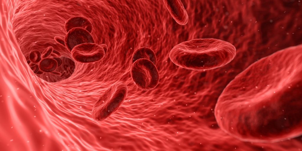
Blood is an essential part of all living organisms. To the naked eye, the blood is just a mass of thick red liquid. In biology classes, we were taught what appears to be this liquid mass has its distinct components, both solid and liquid.
In reality, the liquid portion is the colorless plasma and the red color is as a result of the large percentage of red blood cells. Of course, this can only be recognized and visibly observed with the aid of a high magnifying power microscope.
A compound microscope remains the best option for viewing blood in detail. This high-power microscope is also commonly referred to as the biological microscope.
The blood is made up of four major components: the red blood cells (Erythrocytes), the white blood cells (Leukocytes), the plasma, and the platelets (Thrombocytes).
Having established this, we can proceed to discuss what each of these components looks like under a microscope so you can have better insight of what blood look like under a microscope.
The Red blood cells (Erythrocytes)

Under the microscope, red blood cells are observed as biconcave discs with bright red/pinkish color and no nucleus. The red coloration is an indication of its hemoglobin content. They are the most abundant components of the blood (comprise about 45% of the blood). Erythrocytes have no nucleus or mitochondria at maturity. Their sizes are about 8µm in diameter each.
The red blood cells transport oxygen from the lungs to the other body parts. Because they have no nucleus they can store up more hemoglobin (a protein molecule rich in iron). The cells cannot use the oxygen they transport themselves due to the lack of mitochondria. This is good as it ensures that the other cells receive optimum oxygen. The protein is responsible for binding oxygen atoms. Red blood cells also transport carbon dioxide back to the lungs for expulsion.
Although the human blood has the same composition, they are still not exact matches. Under the microscope, you could notice some particle-like substances that appear to be sprinkled on the surface of the red blood cells. A few of them are known as antigens. The presence of the antigen differentiates a blood type from another. The distinct antigens present in humans are the A, B, and O antigens. Some could have both the A and B antigens. This blood type is known as the AB type.
There is also the Rhesus factor to consider. It is a special type of antigen too. People that possess this antigen are referred to as being Rhesus positive. While those that don’t are Rhesus negative. When it is time for a blood transfusion, only compatible blood is to be transfused. The wrong blood type could be detected as a foreign substance and the immune system of the recipient starts to fight against it. This could result in very critical health issues.
The White Blood cells (Leukocytes)
The white blood cells, although larger, are not as many as the red blood cells. They constitute less than a percent of the blood cells in humans. The White blood cell is a vital component of the immune system which aids the body’s fight against diseases.
They are colorless hence their need to be stained before they can be seen under a microscope. Leukocytes possess a nucleus, mitochondria and have an amoeba-like appearance. They can be further classified into two groups based on their cytoplasm forms.
1. Agranular leukocytes
These are white blood cells with clear cytoplasm. The cytoplasm surrounds the slightly indented dark-stained nucleus. The white blood cells identified under this group are the lymphocytes and the monocytes. Lymphocytes, responsible for the antigen and antibody properties, range in size (usually 6-14µm diameter). They make up more than one-fourth of the entire white blood cell population.
The lymphocytes are best recognized by the round and almost uniform nucleus and little blue cytoplasm usually accompanied with them. Monocytes have a 12-20µm diameter size making them the largest group of white blood cells. Although larger, they make up just 3-8% of the entire white blood cell population.
They have larger cytoplasm and are more irregularly shaped in comparison with the lymphocytes. Monocytes are characterized by a dull grayish cytoplasm.
With the microscope, you may also observe vacuoles as holes in the cytoplasm. The nuclei of monocytes are kidney-shaped.
2. Granular leukocytes
As the name implies, the cytoplasm of this group of white blood cells appears to be filled with granular structures. The white blood cell types that belong to this group include the basophils, eosinophils, as well as neutrophils.
Neutrophils are spherical when viewed under a compound microscope. Generally, the Neutrophilis make up about 70% of the total population of white blood cells. A single one is about 12µm in diameter. They are responsible for bacteria ingestion. They can be easily identified with their lobed nucleus and many light pink/bluish purple granules. The cytoplasm in a healthy individual has light pink coloration. It changes to a darker color in an individual with an infection. This phenomenon is referred to as toxic granulation.
The nucleus of the neutrophils is dark stained under microscope. And its lobes size also varies from 2-5 unevenly sized lobes. In abnormal cases, the nucleus in neutrophils may have more than 6 lobes (e.g. in someone suffering from a special case of anemia).
With a horse-shoe like nucleus, Eosinophil also appears spherical under microscope. It has red (or orange) colored granules.sizes as Neutrophils. It releases a protein that kills large bacteria that are too big to be ingested. The nuclear lobes are relatively more round and uniform in size. The number of Eosinophils usually increases when the host has an allergy reaction or is infected with a parasite.
Basophils, the white blood cells with the lowest occurrence, contain deep blue-purple granules in their cytoplasm. The nucleus of a basophil is bilobed. The basophils are the cells that cause allergic reactions.
An abnormality in the usual volume of white blood cells in the body could be an indication of infections, allergies, or even Leukemia.
Platelets (Thrombocytes)
Platelets are the component of the blood in charge of blood clotting. They help to reduce blood loss when a cut is made to a body part. They have no nucleus. They are small cell particles characterized by many vesicles and are not bigger than 4µm diameter.
They can be observed as spots between the red blood cells under a microscope. And they usually appear like a small plate under a microscope.
Someone with too little amount of platelet suffers from a condition referred to as thrombocytopenia. The condition can cause the patient to bleed out excessively after an injury, cut, or even surgery.
A situation in which there are excess platelets in someone’s blood is termed as thrombocytosis. It causes a lot of unnecessary blood clots and may even end up blocking important blood vessels. The good news is that thrombocytosis is usually a temporary reaction to trauma and goes away by itself. Drugs, alcohol, diseases, and certain food could affect the number and quality of platelets in the blood.
Plasma
The real liquid component of the blood is the plasma and is colorless. It is the main blood component and makes up around 55% of the human blood. Plasma contains a lot of water, proteins, ions, nutrients, and even wastes. It can be observed in isolation with the aid of a centrifuge. The centrifuge spins the blood at a very fast rate with the aim of separation. At the end of the spinning process, the less dense plasma floats on top above the denser cells and cell fragments
How does an unhealthy blood cell look under the microscope
Anemia is a common blood disease. It is conditions in which the red blood cell are too few which reduces the supply of oxygen to the necessary areas of the body. Sickle cell anemia is a special anemic condition.
As observed under a microscope, the red blood cells are not only a few alone. Some of them also have a “C” or sickle shape instead of the regular uniform rounded shapes.
Malaria is another deadly disease. The test for this disease in the blood is normally carried out with the Giemsa stain as a standard. Malaria parasites can be seen as small ring-shaped particles on the red blood cells.
Does animal blood look different from human blood under the microscope?
Many animals have the same blood composition as humans. Human blood and the blood of other animals have same appearance to the naked eye. But there are many differences in their composition.
Hemoglobin responsible for the red blood collection is absent in some animals, especially in invertebrates. For example, polychaete worms have Chlorocruorin pigment which is located in the plasma instead. This pigment can be identified with its green color. The skinks in New Guinea have green blood as a result of the biliverdin pigment in their cells. Octopuses have blue blood because they have hemocyanin instead of the more commonly known hemoglobin. The former attracts copper and turns blue when oxygenated. Crocodile Icefish have colorless blood.
Apart from the general blood color, animals whose blood resembles that of humans have slight differences too. This can be observed in detail with a biological microscope.
Although the red blood cells function the same way in all animals, they are not the same. In humans, the red blood cells are like doughnuts without the characteristic hole in the middle.
But in animals, the shape varies. Some have more rounded red blood cells, while in others; they could be more oval-shaped. Red blood cells in many birds, snakes, and crocodiles are nucleated in opposition to the enucleated ones in humans. In cases of regenerative anemia, the red blood cells in certain animals could be nucleated. Examples of such animals are dogs and cats.
A comparison between the blood of a frog and that of a human with a microscope
The blood of both living organisms looks very alike. They both possess plasma, red and white blood cells. The red blood cells in the frog’s sample appear to be more elliptical in contrast to the well-rounded ones in humans. A dark spot can be observed in the frog’s red blood cells as the nucleus. This is not the case in human red blood cells.
The white blood cells in both organisms have almost the same features. They both stain differently from the erythrocytes and have nuclei. Despite being present in the human blood sample, platelets are absent in the frog’s blood sample.
What is a Blood Smear
A blood smear test is carried out to detect abnormalities of the blood component with a microscope. The unusual appearance of the shape, size, and color of the blood components can be easily noticed with a blood smear test. Too many or too few platelets, red/white blood cells can be noted too. This aids in the right diagnosis of blood conditions.
Apart from anemia, red blood cell diseases could also include hemolytic rustic syndrome and polycythemia rubra vera. The syndrome can be caused by a digestive system infection. The other condition results when there are too many red blood cells in the body.
Diseases that can be related to irregularities with the white blood cells are hepatitis C, HIV, parasitic and fungal infections. Liver or kidney diseases, hypothyroidism, are other conditions that could be indicated by a blood smear.
How to examine blood cells under a Microscope
For proper examination, the following materials are needed:
- A compound light microscope,
- Slides
- Coverslips
- A suitable stain (e.g Leishman’s stain),
- Oil immersion
- A blood sample, and
- A dropper
With the dropper, add a drop of blood to a slide and then use the coverslip to cover it. The slide should be stained to clearly show the cell features. Mount the slide on the stage of the compound microscope.
Start your observation with an objective lens of low power (100x), before you proceed to higher magnifications. Make use of the oil immersion with the high magnification objectives. Do not forget to adjust the lighting and the focus knobs to get a clear image of the blood sample.
With this, you should be able to see numerous red blood cells and fewer white blood cells.
